A team of Canadian researchers recently undertook a study for which they examined using ultrasound-assisted brace casting in patients with adolescent idiopathic scoliosis (AIS). They concluded that real-time 3D ultrasound can help orthotists select the pad pressure and location that provides optimal in-brace spinal correction for this patient population, and that this radiation-free method also reduced the average number of radiographs per subject taken prior to final brace implementation. Their study was published online in the April 11 issue of the open-access, online journal Scoliosis.
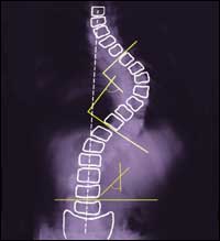
The pilot study included 26 patients with AIS. The control group included 17 participants (two boys and 15 girls at age 13.2 ± 1.5 years) for whom the traditional plaster cast and molded method, with the assistance of the Providence brace system, was used to design their braces. The major out-of-brace Cobb angle for the control group was 32 ± 7 degrees. The average in-brace major Cobb angles at the first in-brace follow-up clinic and at the final accepted follow-up clinic were 22 ± 14 degrees and 14 ± 11 degrees, respectively. The average in-brace Cobb corrections were 31 ± 29 percent at first attempt and 56 ± 27 percent after final adjustments.
For the intervention group of nine participants (two boys and seven girls at age 13.3 ± 1.4 years), in addition to the standard of care, a medical 3D ultrasound system, a custom pressure measurement system and in-house software were used to select pad placement and pressure levels. The orthotist used a custom standing Providence brace design system to apply pressures against each intervention participant’s torso; the applied pad pressures were recorded, and a real-time ultrasound spinal image was displayed. Cobb angle measurements from the baseline and the assessment scan were performed. The orthotist then decided if an adjustment was needed in terms of altering the pad locations and pressure levels. The procedures were repeated until the orthotist attained the best simulated in-brace correction configuration to cast the brace. The major out-of-brace Cobb angle from the radiographs prior to bracing was 29 ± 8 degrees. The average final in-brace Cobb angle was 11 ± 13 degrees, which was a 51 ± 21 percent in-brace correction.
In the control group, 47 percent of the subjects needed a total of 16 brace adjustments after initial fabrication, requiring a total of 33 in-brace radiographs. For the intervention group, the orthotist tried additional configurations in 78 percent of the participants. Among those seven cases, five showed better stimulated in-brace corrections that were subsequently used to cast the brace. As a result, only one intervention subject required a minor adjustment after initial fabrication. The total number of in-brace radiographs in the intervention group was ten.