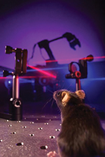
The optical BMI allows bidirectional communication with the brain. While a robotic arm is controlled by neuronal activity recorded with optical imaging (red laser), the position of the arm is fed back to the brain via optical microstimulation (blue laser). Photograph courtesy of Daniel Huber and UNIGE.
Neuroscientists at the University of Geneva (UNIGE), Switzerland, set out to determine whether it was possible to transmit an artificial sensation of prosthetic movement to the brain by stimulating neural activity in the cortex. They discovered that not only was it possible, but that the underlying learning process occurs rapidly. These findings were obtained by using modern imaging and optical stimulation tools, offering an alternative to the classical electrode approach to brain-machine interfaces (BMIs). An article describing their work was published February 22 in the journal Neuron.
Replacing a lost limb with a robotic prosthesis is the subject of much research, with many BMIs operated by relying largely on visual perception, so the flow of information between the brain and the prosthesis is unidirectional. However, movement perception is not based solely on vision but mostly on the sensation of where the limb is located in space. “We have therefore asked whether it was possible to establish a bidirectional communication in a brain-machine interface: to simultaneously read out neural activity, translate it into prosthetic movement, and reinject sensory feedback of this movement back in the brain,” explained Daniel Huber, PhD, a professor in the Department of Basic Neurosciences of the Faculty of Medicine at UNIGE.
In contrast to invasive approaches using electrodes, Huber’s team specializes in optical techniques for imaging and stimulating brain activity. Using a method called two-photon microscopy, they measure the activity of hundreds of neurons with single-cell resolution. “We wanted to test whether mice could learn to control a neural prosthesis by relying uniquely on an artificial sensory feedback signal,” said Mario Prsa, PhD, a researcher at UNIGE and the first author of the study. “We imaged neural activity in the motor cortex. When the mouse activated a specific neuron, the one chosen for neuroprosthetic control, we simultaneously applied stimulation proportional to this activity to the sensory cortex using blue light.” Neurons of the sensory cortex were rendered photosensitive to this light, allowing them to be activated by a series of optical flashes and thus integrate the artificial sensory feedback signal. The mouse was rewarded upon every above-threshold activation, and 20 minutes later, once the association learned, the mouse was able to more frequently generate the correct neuronal activity.
This means the artificial sensation was not only perceived, but that it was successfully integrated as a feedback of the prosthetic movement. In this manner, the BMI functions bidirectionally. The researchers think that the reason this fabricated sensation is so rapidly assimilated is because feeling the position of our limbs occurs automatically, without much thought and probably reflects fundamental neural circuit mechanisms.
Further, the researchers discovered that the mouse “activated only the one neuron chosen for controlling the prosthetic action, and did not recruit any of the neighboring neurons,” said Huber. “This…reveals that the brain can home in on and specifically control the activity of just one single neuron.”
At present, the neuroscientists at UNIGE are examining how to produce a more efficient sensory feedback. They are currently capable of doing it for a single movement, but are exploring the possibility of providing multiple feedback channels in parallel. This research sets the groundwork for developing a new generation of bidirectional neural prostheses that might allow the user to have more precise movements, feel touched objects, or perceive the necessary force to grasp objects.
Editor’s note: This story was adapted from materials provided by UNIGE.