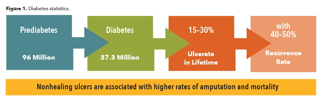
Diabetes mellitus is a disease where the body either does not produce enough insulin or becomes insulin resistant, and there are elevated levels of glucose in the blood stream. With time, high blood sugar has manifold effects including neuropathy due to the deterioration of the peripheral nerves. One of the results is loss of protective sensation. This occurs first where the nerves are finest, in the extremities, leaving them vulnerable to injury and ulceration. Patients with advanced neuropathy often complain of numbness, tingling, or burning in their fingers, hands, toes, or feet. In addition, due to other complications from the disease, when skin breakdown occurs it is slow to heal, taking months or even years. This in turn can begin an expensive cycle of wound infections that may lead to amputation. Diabetes continues to be the number one cause of the approximately 150,000 amputations performed each year in the United States. According to the Centers for Disease Control and Prevention, there are 154,000 hospitalizations associated with diabetic lower-extremity amputations annually, which puts a huge financial burden on the healthcare system (Figure 1).1
Breakdown
Diabetic ulcers occur as a result of several factors including vascular issues, neuropathy, and trauma to the skin. Typically, they first manifest on the feet as full body weight is repetitively transferred with every step. The possibility of a foot ulcer developing is in proportion to the extent of the neuropathy and the magnitude of shear force which can be conceptually represented as follows:
Neuropathy + Shear Force + Time = Ulcer
This simple formula also indicates how a patient with diabetes can be protected. If the advancement of neuropathy can be halted, shear forces held below the threshold of breakdown or the number of damaging cycles limited, ulceration will not occur. However, none of these factors are static; each is liable to change.
Neuropathy
Clearly, neuropathy is the critical component, and limiting it represents the greatest opportunity to avoid the catastrophic cycle of pedal destruction. The goal is to keep patients up, moving, and active, so the single best approach is to focus on the root cause: Prevent neuropathy by educating patients, increasing awareness of the dangers, and promoting a healthy lifestyle.
In response to the critical need for the prevention of foot ulcers, the International Working Group on the Diabetic Foot created an extensive archive of material for healthcare practitioners.2 This includes the initial exam, follow-up, and treatment planning based on the staging. Their foot screening protocol is an excellent guide to quickly evaluate and determine a patient’s risk category. They recommend all patients with diabetes at risk for peripheral neuropathy be tested at a minimum annually using Semmes-Weinstein monofilaments to record touch pressure sensation.
Shear Force
Although pressure is often cited as a causative factor of breakdown, it is more correct to name shear force as the culprit. Pressure can be considered a proxy for shear, but they are different. Pressure is a measure of force applied over an area, and skin can withstand reasonable amounts of static pressure. However, shear is the active force when two adjacent surfaces move over each other. Whereas pressure acts down on a surface, shear is a result of sliding. It is possible to have pressure without shear, but you will not have shear without pressure. Poorly fitted footwear, either too tight or too loose, an incorrect shoe shape, or a low toe box can cause unnecessary rubbing and friction, leading to blisters and eventually ulceration.
Shear force can be dramatically reduced by selecting proper shoes, sized correctly for the larger foot. Footwear for people with diabetes should have minimal seams, a smooth low-friction lining, a wide and high toe box, and removable extra-depth inlay. An ideal shoe supports the foot’s natural shape, has a slightly elevated heel (10-12mm drop), a closure that accommodates mild edema, and full-length total contact insoles. When neuropathy is more advanced, patients benefit from customized insoles or custom orthotics fabricated to redistribute load and protect specific areas of high pressure. In the worst cases, custom-molded shoes may be prescribed.
As friction is a key component in the development of shear, patient skin condition is also crucial. Patients with diabetes often have dry, brittle skin. This is a consequence of poor circulation and glycosylation, an irreversible cross-linking of collagen and keratin that causes a thickening of the skin, tendons, ligaments, and joint capsules. The integrity of dry skin can be maintained using a prescribed moisturizer. Finally, hosiery is an important consideration. White socks that are thin on top with plantar padding, a loose nonconstricting fit, and seamless construction, especially at the toes, are less likely to precipitate a problem. It is also recommended to wear socks that are moisture wicking and breathable to prevent the buildup of bacteria.
Time
The component of time refers to both how long the patient has had neuropathy and how frequently shear force is experienced. Nerve damage, which is cumulative and nonreversible, occurs when blood sugars are out of control, beyond their recommended limit. The first line of defense is maintaining proper glucose and A1C levels.
Mechanical damage occurs when forces are above the threshold for destruction. Reducing the number of repetitions or lessening the friction causing shear limits this potential. Although not ideal, there are ways to curtail the amount of time a patient spends walking. Part of the efficacy of total-contact casts and CROWs, if the patient will wear them, is that they impede mobility. Less steps equal less cyclical loading. Of course, these devices work in other important ways protecting the foot and reducing movement within the device.
The Intrinsic Minus Foot
One consideration that can sometimes occur with advancing neuropathy is the changing presentation of the foot itself. The term “intrinsic minus foot” was introduced in the 1970s by Heinz I. Lippman, MD, to explain the effect of peripheral neuropathy on the distal motor nerves that activate the intrinsic muscles of the foot. Although the full mechanism of the deformity is an involved process, and not entirely agreed upon, the main steps can be summarized as follows:3
- In normal feet, the lumbricals flex the metatarsal phalangeal joints (MPJs) and extend the interphalangeal joints (IPJ) of the lesser toes.
- If they lose their efficacy due to neuropathy, the MPJs extend, and the toes flex, resulting in hammertoes and prominent metatarsal heads.
- The change in position of the bones of the forefoot results in a relative distal migration of the fat pads beneath the metatarsal heads, leaving the plantar foot more vulnerable to areas of localized pressure.
- As the situation progresses, skin callusing begins to appear under the metatarsal heads.
- With time, the atrophying of these muscles can lead to a significant change in the shape of the foot. The “downward” progression of the forefoot in relation to the rearfoot may develop a claw-like appearance.
- Other changes occur simultaneously, and it is sometimes noted that the arch height also increases.
The net effect is a dynamically changing insensate foot that results in areas of plantar pressure and hammertoes. The foot needs to be off-loaded using an accommodative insole and protected with a well-fitting, extra-depth shoe with a high or stretchable woven toe box.
Protecting the Forefoot
Due to a number of reasons, the majority of diabetic foot ulcers occur in the forefoot. Firstly, this is where distal neuropathy begins to attack. Secondly, in terminal stance, the smaller area of the forefoot alone supports full body weight as the limb prepares for propulsion. Finally, in the third rocker, restrictions in joint range of motion can increase ground reaction force (GRF) distally (Figure 2).
The smooth operation of the first MPJ is of immense importance.⁴ During terminal stance, once heel lift begins, the first MPJ is the pivot around which the body rotates. Functional hallux limitus is a restriction that occurs only when the joint is loaded. There is a distinct lack of motion at the first metatarsal head and a perceived locking of the joint. This can lead to a variety of compensations as the body naturally seeks alternate paths to continue forward. One possibility is to externally rotate and abduct the foot so that body weight passes over the side of the foot rather than through its long axis. These patients often have a pinch callus on the medial side of the hallux, a precursor for an ulcer. Inspection of the insole can reveal an absence of wear under the first metatarsal head, and clear indications of pressure under the great toe and sometimes the lesser metatarsals.
Certain orthotic designs help restore some of this lost motion as plantarflexion of the first metatarsal bone aids hallux dorsiflexion. A well-fitting custom foot orthotic will raise the medial longitudinal arch, support the midfoot and maintain the first metatarsal in proper sloped alignment. In addition, accommodations can be made to allow the first metatarsal head to sit lower. A first ray cut out in the orthotic shell slightly drops the first metatarsal head. Likewise, a reverse Morton’s extension provides a channel that the first metatarsal can sit in. A kinetic wedge is a rectangular cut out distal to the orthotic shell that creates a depression for the first metatarsal head. These approaches allow the first metatarsal head to drop down and improve motion at the joint; they also serve to transfer some of the forces laterally.
Equinus is a common foot and ankle condition that is believed to be a leading contributor to foot pathology.⁵ In the case of diabetics with advanced neuropathy, it is strongly linked to the breakdown and ulceration of the forefoot. In simple terms, equinus is the inability of the ankle joint to dorsiflex sufficiently, which inhibits the body’s mass from passing over the plantigrade foot in the second rocker. Although there is no universal standard as to what constitutes equinus, a widely accepted value for static measurement of dorsiflexion at the ankle joint is 10 degrees. Any restriction in motion at the ankle joint, initiating early heel rise, means the insensate forefoot will be bearing body weight longer, prolonging the time that the smaller area of the forefoot must sustain GRF.
The worst effects of equinus can be limited through using heel lifts that reduce the magnitude and duration of GRF acting under the forefoot. Conversely, full-length solid ankle AFOs and CROWs that restrict flexion at the ankle and metatarsal heads also reduce peak pressure under the forefoot by maintaining the foot in midstance. Although they alter gait, they may be a necessary compromise to protect the foot.
Rocker Soles
Rocker soles assist movement in the sagittal plane and lessen pressure under the forefoot. They are an aid to restoring fluid motion and the natural rockers of gait when there is a restriction, whether due to joint limitation or an orthotic device. Benefits include a reduction in GRF and compensation for rigid joints. It is possible to guide motion and disperse pressure by adjusting the angle of the fulcrum, its location, and the pitch of sole.
In one study the biomechanical effects of prefabricated foot orthoses and rocker‐sole footwear were examined in individuals with first MPJ osteoarthritis.⁶ The 102 subjects were randomly assigned the orthotic or a rocker-sole shoe and then wore both sensor motion analysis and in-shoe plantar pressure measurement systems. It was noted that “the rocker-sole footwear resulted in reduced cadence, percentage of the gait cycle spent in stance phase, and sagittal plane hip range of motion.” Both interventions significantly reduced peak pressure under the first MTP joint, and the rocker-sole shoes also reduced peak pressure under the second through fifth MTP joints and heel. They concluded in part, “prefabricated foot orthoses and rocker-sole footwear are effective at reducing peak pressure under the first MTP joint….” Although this study focused on patients with first MPJ osteoarthritis, it demonstrates the effectiveness of rocker soles in redistributing forefoot plantar pressures.

Conclusion
Patients with prediabetes have a blood glucose level that is above normal but not yet high enough to be classified diabetic. This stage is reversible. It remains the single best opportunity to curb the progress of this morbid epidemic. Educating patients and keeping them up and active remains the best medicine. Direct benefits include higher patient quality of life, and significantly lower healthcare costs. If neuropathy can be avoided in the first place, the body will act as its own sentry against shear force and skin breakdown.
Once neuropathy takes hold, vigilance is required to prevent injury by wearing properly fitting protective shoes and socks and reducing pressure from weight bearing activities and ambulation. The feet must be inspected daily for signs of skin irritation, pre-ulcerative callusing, or deformity. As the disease advances, the patient’s biomechanics must be evaluated. It plays an important causative role in generating shear force and, if left unaddressed, healed ulcers are likely to reopen. Orthotic and pedorthic devices can be designed to limit the most destructive effects of shear force.
Séamus Kennedy, BEng (Mech), CPed, FAAOP(A), is president and co-owner of Hersco Ortho Labs, New York. He can be contacted at [email protected] or by visiting hersco.com.
References
- https://www.cdc.gov/diabetes/data/statistics-report/index.html
- https://iwgdfguidelines.org/guidelines-2023/all-guidelines-2023
- Bernstein, R. K. 2003. Physical signs of the intrinsic minus foot. Diabetes Care 26(6):1945-46; PubMed:12766145.
- Kennedy, S. 2014. Hallux limitus and the vital operation of the first MPJ. The O&P EDGE 15(5):60-2.
- Kennedy, S. 2013. Equinus. The O&P EDGE 12(4):66-70.
- Menz, H. B., et al. 2016. Biomechanical effects of prefabricated foot orthoses and rocker‐sole footwear in individuals with 1st metatarsophalangeal joint osteoarthritis. Arthritis Care & Research 68(5): 603-11.