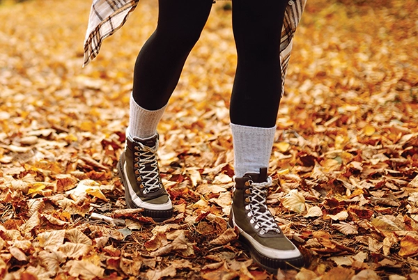
Fall is a popular time for hiking and outdoor adventures. While trails can offer great views of nature, they also have many obstacles that can injure you on your journey. Tree roots and rocks are obvious obstacles, as are slippery slopes on the inclines and declines from loose gravel, dirt, or mud, but when these also become buried under leaves, you may find yourself an unsuspecting victim of their hazards. Despite these impediments, the Werner Herzog quote, “The world reveals itself to those who travel on foot,” epitomizes the allure for many hiking enthusiasts who enjoy the calm crisp sounds of fall, the colors of the leaves, and the fresh smell of the air.
In a previous article, I cited numerous methods for blister prevention that are essential to consider when hiking.1 When we consider our patient population, many conditions combine with the aforementioned challenges: spasticity, muscles firing when they are not supposed to be firing (such as at the end of stance phase when dorsiflexors are firing but the plantarflexors are still firing and opposing the motion, making it more difficult to clear the terrain), muscle imbalances, issues with muscle coordination, foot deformities, forefoot varus/valgus, and hindfoot valgus/varus. So the orthotist’s task is to provide as many strategies and tools as possible to control those factors, helping patients achieve balance by keeping their center of gravity in their base of support, and providing the best base of support possible.
In this two-part article, we will take a closer look at treating or preventing foot and ankle complex injuries on the uneven terrain of the outdoors to help our patients build an army of preventive and accommodative measures to allow them to enjoy the activity of hiking.
Whether we are exploring normal or pathological gait, it is paramount that we first focus globally on the three rockers of walking that prevent more proximal compensations, and what it means for our patients when one or more of the rockers are absent due to anatomical issues. With a normal three-rocker system of walking as depicted in Figure 1, the heel hits the ground, the ankle then plantarflexes to accept the load and to get plantigrade, then the tibia rolls over the foot, and finally the ankle plantarflexes again to a more digitigrade position in preparation for pushing off and going into swing phase. It can be tough enough to escape injury if you are proceeding through all three rocker stages, but if an individual is afflicted with limitations that impact these three rockers, such as limited joint range of motions, spastic hypertonicity, dystonia, or cocontraction of muscles, then extra special consideration is needed to prevent injury. In determining the best orthotic approach, it is important to consider whether the goal is prevention of the pathologies or accommodating for existing pathologies without limiting patients’ freedoms on the trails.
Part one of this article series includes strategies to address pathologies affecting the ankles.
A typical and common presentation of patients with hemiplegia from disorders such as a cerebral vascular accident, cerebral palsy, etc. favors an equinovarus position of the ankle that pre-positions the foot and ankle at heel strike into a forefoot strike first instead as seen in Figure 2. This common presentation in the hemiplegic population can be from contracture or spasticity or both of the gastroc soleus muscles/tendon. This pre-position of landing at initial strike tends to be on the fifth metatarsal head thereby creating a very poor, small base of support for accepting the load for weight bearing, and the patient tries to maintain the center of mass over this small base of support as the contralateral limb begins to come off the ground and go into swing phase on that side. Orthotic intervention becomes essential to prevent coronal and transverse plane injury especially on uneven terrain and declines/inclines that can lead to increased trouble with stabilizing the ankle and preventing strain, sprain, or fracture.
Ankle Sprains
Ankle sprains are a common occurrence in sporting activities and hiking through an overstretching of the ankle ligaments where the collagen fibers are partially or completely disrupted.2 A lateral ankle sprain is more common than medial because of the medial’s strong deltoid ligament.2 The anterior talofibular ligament and calcaneofibular ligaments on the lateral side are more likely to become sprained through a supination torque.3 This occurs with plantarflexion and landing with the foot incorrectly positioned in inversion.3 Once a person sprains an ankle, he or she is more likely to reinjure it.4
Keeping in mind, as previously mentioned, that people with hemiplegia are at increased risk for spraining due to the pre-positioned equinovarus ankle at initial contact, the goal is to prevent this equinovarus from initiating or, in cases of fixed deformity, to do everything possible for the center of gravity in the base of support to equal balance. Individuals with Charcot-Marie-Tooth disease, tight or short Achilles tendons, or those with proprioceptive defects may be more susceptible to sprains as well. Some methods to prevent injury or reinjury include taping, braces, and neuromuscular training to support the ligaments.4 Supportive shoes that have a wide base of support with a stiffer sole can also help reduce the possibility of a sprain. Lateral buttresses or extensions can help accommodate fixed deformities of supination by allowing the center of gravity to fall within an extended base of support in the coronal plane as presented in Figure 3. If the supination is flexible and correctable to a subtalar neutral, then the use of wedging can have a significant impact as it changes the level of the earth under patients’ feet. A Coleman Block Test (Figure 4), in which various wedges are tested on the lateral forefoot and/or hindfoot to determine the most effective way to get the subtalar joint to a neutral position, provides great guidance and foundation for support. In patients who present with supination, prophylactic taping and bracing could be done to prevent initial injury and reinjury. Keep in mind that to control a joint you must cross the joint and use a three-point pressure system, so it is important to support the ankle with an orthosis that extends proximally above the ankle for increased stability on uneven terrain (Figure 5). Orthoses ranging from off-the-shelf ankle-stabilizing braces to custom-molded designs are an essential tool to manage the risk of sprains on uneven ground. Remembering these are triplanar deformities, a custom-molded cast is often needed to prevent skin-tissue injuries from the orthosis, especially if there are bony deformities, contractures, or spasticity/dystonia involvement.
Foot Drop
Sagittal plane mechanics also greatly impact our patients and need consideration as well. In normal gait, we shorten our limb to allow for ground clearance in swing phase by flexing the hip, flexing the knee, and dorsiflexing the ankle. If one of these is impacted, then the others must compensate. In the case of foot drop, more knee flexion or hip hiking, and hip circumduction or increased hip flexion must happen to compensate to clear the ground. At mid-swing, the contralateral leg is in momentum to transition into swing as the ipsilateral is on its way to accepting load, so a foot drop that catches the ground tends to lead to a fall, as there is no longer a stable leg on the ground. Injury prevention is easily accomplished in patients with foot drop through assisting dorsiflexion in swing phase. The challenge is to accomplish this dorsiflexion assistance and still maintain the three rockers of walking.
Various orthoses that allow for free plantarflexion and dorsiflexion assist can accomplish these goals, and recent advances in preimpregnated carbon fibers have allowed a concept of resisting and assisting motions in the material resilience itself. For instance looking again at Figure 1 and also examples shown in Figure 6, in a carbon fiber strut, as the ankle plantarflexes, the strut is working to right itself back to neutral and gives a pull toward dorsiflexion. As the ankle progresses into the second rocker, and the ankle dorsiflexes bringing the body over the foot, the strut wants to pull back out of dorsiflexion to its neutral position, assisting mid- to late-stance extension. And finally, in the last stance phase of gait, the ankle plantarflexes and the strut screams “put me back to neutral,” thereby propelling the foot as well to a neutral position in swing, allowing it to clear and also hit a good normal heel strike again to start the cycle over.
Achilles Tendinopathy
Achilles tendinopathy is a common overuse injury for active individuals, especially in sports involving running and jumping.5 Pain, swelling, and loss of function occur in the Achilles tendon. The injured tendon progresses into tendinopathy when there is continued overloading of the tendon without time to heal.5 The Achilles tendon can be overloaded through compressive forces in ankle dorsiflexion through activities such as running, hiking, or traversing uneven terrain. Haglund’s deformity, or posterior calcaneus protuberance, may also be involved in producing pain.5 Additional risk factors include decreased plantarflexor strength, limited ankle dorsiflexion or subtalar joint motion, and excess pronation. Early treatment is important to prevent the injury from progressing. Treatment or prevention options include strength exercises, taping, and orthotics. If a patient is having tenderness in the Achilles area, it may be beneficial for an orthosis to help resist dorsiflexion.
When considering an orthotic intervention, keep in mind that if a patient with spastic hypertonicity loads the foot and then transitions into tibia advancement, especially on a downhill, a spastic gastroc soleus firing could cause large strains and stresses to the Achilles tendon. Stopping dorsiflexion is an option but resisting the sagittal motion would be better. In other words, allowing for motion but resisting it—picture someone behind the hiker with a rope pulling on the tibia, slowing it down and taking stress off it. The key here, and in all injury prevention, is to listen to the body; when the “check engine lights” come on, when you feel pain, pay attention.1
Calcaneal Spurs
Calcaneal spurs are cartilaginous formations that develop on the plantar or dorsal surface of the calcaneus.6 Plantar spurs emerge on the back of the heel near the Achilles tendon insertion and are commonly called Haglund’s deformity or pump bumps.7 Dorsal spurs form on the bottom of the heel close to where the plantar fascia originates.6 The cause of these spurs is not completely known, but it is connected to conditions such as arthritis, high body mass index, plantar fasciitis, pes cavus, and pes planus.6 These calcaneal spurs can cause pain when ambulating, which makes it a challenge for athletes and hikers. There are multiple conservative methods to manage pain and prevent irritation of the calcaneal spurs. For those with dorsal calcaneal spurs, a heel height of 2-3cm can be added using a heel wedge to transfer some of the weight from the heel to the midfoot and forefoot.8 When buying hiking shoes or trail runners, take note of shoes with a higher heel than forefoot versus shoes that are completely flat. Custom foot orthoses and thick, soft padding such as silicone around the spur can also reduce pain.
People with cerebral palsy often have flat feet, so custom foot orthoses and shoes that do not irritate the heel can prevent these spurs from developing. Stretching the gastroc soleus and fascia can further reduce irritation of plantar spurs and dorsal spurs respectively.6 Patients with diplegia with spasticity often have tight or shortened Achilles tendons, so stretching can help stop the spurs from forming or reduce symptoms if already present by preventing the compensatory gait pattern of early and excessive heel rise when the Achilles is tight.
From personal experience with plantar calcaneal spurs, extra padding around the counter of the shoe is more comfortable than stiff and unpadded counters such as rubber boots. If a patient is currently wearing a thermoplastic AFO, applying a heel bumper or cutout with additional padding can help prevent these extra pressures as well. ShearBan applied on the shoe or orthosis will also reduce friction.1 For patients afflicted with a heel spur under the heel, it is essential to prevent any pronation to relieve stresses or strains in that area, and in the pes cavus or supinated foot the goal is to redistribute the weight bearing throughout the foot and lessen it at the heel spur site by bringing the ground up to reach every aspect of the foot, relieving pressure in that specific area.
Midfoot Collapse
In patients with diplegia, pronation is a common presentation. One component that needs special consideration is the midfoot collapse aspect. We like to describe this midfoot collapse scenario with our patients as a rock and bridge scenario. Is the rock simply dropping from the bridge into the water below or is it being thrown and propelled by an external force towards that water? In other words, is the arch just collapsing in a flaccid scenario, or is it being forced down by a limitation, for instance by the gastroc soleus range of motion (ROM)? We must understand a key component of stance phase of gait here and in numerous scenarios with our patients. The body must propel over the foot, and it will find every way to do it through the path of least resistance. If the ankle ROM does not allow for the tibia to roll over the foot, then the path of least resistance is often midfoot collapse through the talonavicular and calcaneocuboid joints (midtarsal joints) and a new hinge is formed as a result.
As orthotists, our goal is to prevent joint deformities. So for patients with pronated feet, we must support their arches for them to successfully navigate hiking on hills. Foot orthoses provide a simple solution for the flaccid presentation of midfoot collapse by providing a support beam structure to the falling bridge caused by conditions such as plantar fasciitis and posterior tibial tendonitis. The orthosis prevents joint deformity and soft tissue injuries through arch support. In the limited-range scenario, a good arch support with a rocker sole may be helpful to relieve the stress on this forced midfoot collapse—like the rock being thrown rather than dropped from the bridge. A focus with physical therapy and/or bracing is to improve ROM and as a result to allow for the motion to be restored from the ankle as well to decrease the magnitude of these forces. If you notice that the ankle ROM does not come to 90 degrees, then a heel wedge may be essential in the armament as well to bring the ground up to meet the foot and ensure then that the tibia angle to the earth is 90 degrees.
Posterior Tibial Tendonitis
The posterior tibial tendon (PTT) is a large tendon that runs anterior to the medial ankle and acts as the main medial longitudinal arch stabilizer and produces midfoot inversion and ankle plantarflexion.9 PTT dysfunction (PTTD) or rupture is the main cause of acquired flatfoot in adults, and it progresses into other deformities such as forefoot abduction, calcaneovalgus, or fixed varus-supination deformity.9 The cause of PTTD is unknown, but it is a progressive condition that starts with tendinopathy of the posterior tibialis and can progress to subtalar arthritis and deformity of the ankle and foot.10
Patients with diplegia are particularly susceptible to PTTD due to their hyperpronated positions and related high stresses and stretching of the PTT complex, especially at the early stages of PTTD. Orthotic devices including foot orthoses, UCBLs, Arizona Braces, and AFOs are used to relieve the PTT and eliminate forefoot pronation.10 These orthoses include arch support and a medial forefoot wedge. Exercises to strengthen and stretch the PTT include resisted foot adduction and calf stretching.11 The goal is to help assist the function of the PTT and to prevent the cycle from development of pronation to PTTD or vice versa, leading to more pronation and to an increased stage of PTTD. Often, free motion in the sagittal plane but coronal and transverse limitations need to be aggressively controlled, which could involve the use of an AFO to help the PTT with its function of helping to prevent pronation on the trails if there is any suspect.
The authors’ objective in part 1 is to help O&P patients enjoy the great outdoors and its many pleasures through hiking. We aim to provide interventions for common pathologies and help patients meet the goals of being able to achieve balance physically and mentally with the security of preventive and accommodative measures. In part 2, we will continue with a focus on working with the many foot deformities and how to help patients hit the trails with those issues. Happy hiking!
Keith M Smith, CO/L, FAAOP, is a practitioner with the Orthotic and Prosthetic Lab, Missouri. He can be contacted at [email protected].
Brooke Merryman, MSOP, is a resident with the Orthotic and Prosthetic Lab, Missouri. She can be reached at [email protected].
References
- Smith, K. 2023. An orthotist’s perspective on blister prevention from hiking the Camino de Santiago Trail. The O&P EDGE 22(6):28-36.
- Oscar, L. W-H., L. Tun-Hing, and C. Kai-Ming. 2011. The epidemiology of ankle sprain during hiking in uniformed groups. Journal of Orthopaedics, Trauma and Rehabilitation 5:10-6.
- Wright, I. C., R. R. Neptune, A. J. van den Bogert, and B. M. Nigg. 2000. The influence of foot positioning on ankle sprains. Journal of Biomechanics 33(5):513-9.
- Verhagen, E. A., and K. Bay. 2010. Optimising ankle sprain prevention: a critical review and practical appraisal of the literature. British Journal of Sports Medicine 44(15):1082-8.
- Silbernagel, K. G., S. Hanlon, and A. Sprague. 2020. Current clinical concepts: Conservative management of Achilles tendinopathy. Journal of Athletic Training 55(5):438-447.
- Velagala, V. R., N. R. Velagala, T. Kumar, A. Singh, and A. M. Mehendale. 2022. Calcaneal spurs: A potentially debilitating disorder. Cureus 14(8):e28497.
- Choo, Y. J., C. H. Park, and M. C. Chang. 2020. Rearfoot disorders and conservative treatment: A narrative review. Annals of Palliative Medicine 9(5):3546-3552.
- Wibowo, D. B., R. Harahap, A. Widodo, G. D. Haryadi, and M. Ariyanto. 2017. The effectiveness of raising the heel height of shoes to reduce heel pain in patients with calcaneal spurs. Journal of Physical Therapy Science (12):2068-2074.
- Guelfi, M., A. Pantalone, R. M. Mirapeix, D. Vanni, F. G. Usuelli, M. Guelfi, and V. Salini. 2017. Anatomy, pathophysiology, and classification of posterior tibial tendon dysfunction. European Review for Medical and Pharmacological Sciences (1):13-19.
- Ling, S. K., and T. H. Lui. 2017. Posterior tibial tendon dysfunction: An overview. Open Orthopaedics Journal 11:714-723.
- Kulig, K., S. F. Reischl, A. B. Pomrantz, J. M. Burnfield, S. Mais-Requejo, D. B. Thordarson, and R. W. Smith. 2009. Nonsurgical management of posterior tibial tendon dysfunction with orthoses and resistive exercise: A randomized controlled trial. Physical Therapy (1):26-37.
Top photograph: maxbelchenko/stock.adobe.com
Photographs courtesy of Keith Smith and Brooke Merryman.