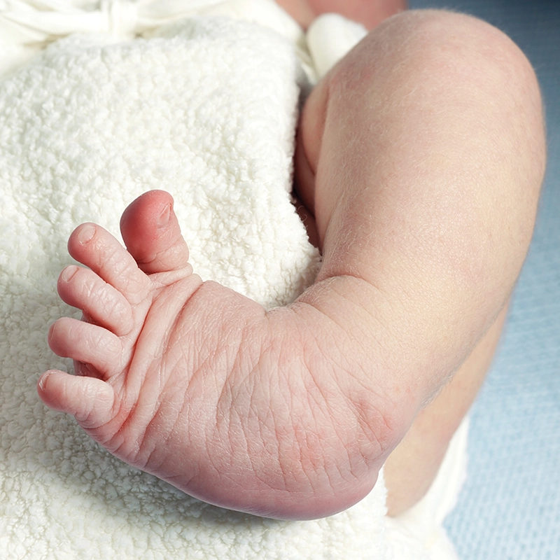
Talipes equinovarus, or clubfoot, is a common congenital disorder of the foot. The Latin term talipes is derived from the conjunction of talus, the ankle bone, with pes, the foot. While there are several congenital deformities involving the talus, the talipes equinovarus foot presents in an inverted and plantarflexed position.
It is best considered a complex, multiplanar deformity of the lower limb as it affects more than just the foot. The calcaneus is rotated under the talus, the rearfoot is inverted, and the forefoot is adducted and in equinus. There may also be muscle imbalances, soft tissue contractures, and a tight Achilles tendon. Left untreated, the deformity will increase, the foot will become more rigid, and ultimately, gait will be affected.
Prevalence
In the literature, talipes equinovarus is reported as approximately one per 1,000 births, although there is plenty of variation across populations. Hawaiians and Pacific Islanders have an incidence as high as seven per 1,000, while Asians have a much lower range of only 0.4 per 1,000. It affects boys at twice the rate compared to girls, half of which are bilateral. The majority of cases, approximately 80 percent, are considered idiopathic, and the remainder are linked to neuromuscular or genetic disorders.1 Family history also plays a role: Parents born with clubfoot are more likely to have a child with the condition, and even more so if there is a sibling with clubfoot.
Signs may be noted in prenatal ultrasounds, and physicians examine for clubfoot at birth. The affected foot, or feet, are inverted, lacking eversion and dorsiflexion. There is often a signature crease at the posterior ankle and medial arch. Early identification is important as treatment should be initiated almost immediately. It begins with serial casting or manipulation of the foot and ankle and then proceeds to bracing and shoe therapy.
The Ponseti Method
Once diagnosed, clubfoot should be treated as soon after birth as possible while the joint structures remain highly mobile. The Ponseti Method is an extremely successful, conservative approach to addressing clubfoot in infants and young children. It was developed by Ignacio Ponseti, MD, in the mid-20th century as an alternative to corrective surgery. Interestingly, it only became widely adopted in the 1990s after he published a book, and word spread across online forums among concerned parents.2 The pioneering work of Ponseti and colleagues at the University of Iowa’s Carver College of Medicine has helped generations of patients worldwide. It has been especially potent in developing countries where this relatively simple method, once properly understood and learned, is easily applied at low cost.
The underlying complex anatomy of a clubfoot can be characterized by a useful acronym that highlights the major structural components of cavus, adductus, varus, and equinus (CAVE). These occur due to combinations of tightness in various intrinsic muscles, the long flexor tendons, the gastrocnemius, the posterior tibial tendon, the anterior tibial tendon, and the plantar fascia that leaves the bones malaligned.3 To achieve a beneficial outcome from casting, each of these components must be addressed.
The goal of casting is to rotate the foot around the talus and redirect growth. Prior to casting, the joint is manipulated to reduce soft tissue contracture. Then the limb is cast in a fixed position from the toe to the upper leg. It is important to proceed with correction in the proper sequence, from distal to proximal. Casting should reduce the adducted forefoot first, then the inverted varus of the rearfoot, and finally the equinus.
If the correction is done out of turn, the foot may develop other malformations. For example, the temptation to correct the equinus first, which is a visible aspect of the deformity, must be avoided, as it can result in creating a rocker-bottom foot.
Casting is performed approximately weekly and continues for several months. Proper alignment can be confirmed by radiographs. In addition to casting, many cases require a percutaneous tenotomy of the Achilles tendon to release some of the plantarflexion contracture. The Ponseti Method is considered the gold standard for early treatment with documented success rates greater than 90 percent.
The French Functional Method is a second conservative technique of intense physical therapy and manipulation. It relies on daily massaging of the foot and lower limb to release the muscles and joints to correct foot position. Although both methods are documented as being very successful, the ultimate outcome for both depends on following a strict bracing protocol after initial correction has been achieved.
Orthotic Bracing
Recurrence rates of clubfoot are high, and even after successful serial casting, it is notoriously difficult to maintain the correction in a growing child. If the patient is under two years of age, a second round of casting may be initiated. Part of the problem is that clear success of early manipulation, which can fully correct the deformity, leads to complacency. To prevent relapse, it is vital that the parents adhere to bracing once correction is achieved.
The first transition is to use an abduction bar along with orthoses that substitute for the cast. Conventional approaches such as the Denis Browne splint and several modern dynamic abduction braces have been incorporated into modular systems that allow the clinician to adapt the treatment as the child develops. These bar-bracing systems incorporate AFOs or footwear that are easily attached. They work by simultaneously externally rotating the foot, blocking forefoot adduction, and preventing ankle plantarflexion.
Initially it is expected the infant wears the apparatus more than 22 hours each day for several months. As the young patient approaches walking, there will be less wear time, but the brace should still be applied while napping and sleeping at night. Orthosis wear may continue for up to five years until the child rejects the treatment.
Shoes
Traditionally, once the toddler was walking, leather footwear was used to maintain the correction. Stiff, high-top shoes with reverse lasts and secure closures held the foot in an optimal position.5 The benefits could be further enhanced with the application of internal pads and external wedging. Nowadays, practitioners can use specifically designed AFOs to achieve the same results. Features may include plantarflexion stops and forefoot dorsiflexing wedges.
There are several pediatric shoe brands that offer a variety of roomy, straight-lasted footwear that accommodate AFOs. They are less orthopedic looking and sportier. Parents should know that the affected foot may be a different size, and this needs to be considered when sizing shoes.
Once they begin walking, children’s feet should be inspected for signs of pressure spots or blisters to ensure each shoe is sized correctly. Seating the heel properly in the brace is very important to prevent a permanent Achilles contracture or creating another deformity due to a break of the midfoot. Children may be prescribed AFOs with long walls to control foot position in the transverse plane, and multiple straps to hold the foot in place.
Older Patients
It is quite common to see young adults and older patients with some residual characteristics of clubfoot. Typically, they will have a high-arched, stiff pes cavus type foot with a thinner calf—different than the contralateral side. It is not unusual that the feet are two distinctly different sizes. There may be some combination of metatarsus adductus, a visible prominence at the base of the fifth metatarsal, ankle equinus, metatarsalgia, and some symptoms of arthritis. A biomechanical evaluation may reveal unilateral limited subtalar joint range of motion, reduced calf musculature, internal tibial torsion, and a limb length discrepancy.
Foot orthotics can be designed to alleviate bony protrusions, especially the base of the fifth metatarsal, and to cushion the metatarsal heads. Materials such as EVAs and cork are best suited to accommodate the foot shape. Extra padding from Poron, foam rubber cushions, and Spenco top covers provide welcome shock absorption. In younger patients, a functional rigid gait plate may be prescribed to address in-toe gait. A lift can be added to the orthotic and/or the shoe to account for a leg length difference.
If the foot size difference is not too great, high-top shoes can help secure the smaller foot in place and allow room for an orthotic. Beyond a difference of two to three shoe sizes, the patient may use a toe filler or have to consider buying two different size shoes. Restrictions in ankle motion can be augmented with rocker soles, which should usually be applied bilaterally to maintain symmetry in gait.
Surgery
When conservative measures fail or the deformity is severe, surgery may be necessary to address the situation. Surgery on children is now rare and generally only considered after serial casting and bracing have not produced the desired results. There are a variety of bone osteotomies, foot column lengthening procedures, and tendon transfers that surgeons will consider to correct the foot.
Conclusion
Talipes equinovarus is a common congenital birth disorder that is well understood. Although it is a complex deformity of the lower limb, it can be successfully treated with conservative casting and manipulation. However, recurrence rates in the rapidly developing young limb are extremely high. To prevent relapse, aggressive orthotic and footwear therapy are necessary along with parent education. Older patients with mild signs of residual clubfoot often benefit from foot orthotics that incorporate depressions, lifts, and pads to accommodate their feet.
The author gratefully acknowledges the help and contribution of Caryn Meth-Gottfried, CPO, in reviewing this article.
Séamus Kennedy, BEng (Mech), CPed, FAAOP(A), is president and co-owner of Hersco Ortho Labs, New York. He can be contacted at seamus@hersco.com or by visiting hersco.com.
References
- Williams, M. 2022. A guide to treating pediatric clubfoot. Podiatry Today 35(12).
- Ponseti, I. Congenital Clubfoot: Fundamentals of Treatment. Oxford University Press, 1996.
- Kleeman, T. J. The Active Foot: From First Step to Last–Clubfoot. The Pedorthic Footcare Association webinar series, June 4, 2025.
- Richards, B. S., S. Faulks, and K. E. Rathjen, et al. 2008. A comparison of two nonoperative methods of idiopathic clubfoot correction: the Ponseti method and the French functional (physiotherapy) method. Journal of Bone & Joint Surgery 90(11):2313-21.
- Caselli, M. A. 2004. The role of shoe therapy in the management of pediatric congenital clubfoot. Podiatry Management 23(8):125-31.
Image credit: AGLPhotoproductions/istock.com.