
Outcomes for patients with plagiocephaly are typically reported by recording the initial level of oblique asymmetry present and the amount of correction afforded by a cranial remolding orthosis (CRO). 1,3-6 Cranial vault asymmetry (CVA) is used by many insurance companies as a criterion for medical necessity, so it is convenient to reference clinically for insurance approval and use as an outcome measure. These point-to-point measurements are limited: They don’t represent the entire 3D aspect of an abnormally shaped head and often do not reflect the primary concern of the parents/caregivers.2, 7-10 A CRO may improve the CVA below 5mm, but substantial asymmetry that is not accounted for by the CVA measurements may still be present.
Three-dimensional correction can be visible using widely available surface scanning, which offers a qualitative outcome for parents.8-10 However, there is not an easy quantitative manner to represent this aspect of plagiocephaly (or other 3D aspects of plagiocephaly), so it is rarely reported on in the literature. This article presents two separate cases where the lateral and anterior asymmetry was the primary concern of the parents and subsequently became the primary goal of treatment.
In both cases, the patients presented with a similar CVA (approximately 16mm difference) and right posterior and left anterior flattening. Both also had significant anterior asymmetry and obvious asymmetry in the frontal plane.
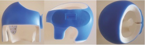
In both cases, a custom Boston Band (Boston Orthotics & Prosthetics, Avon, Massachusetts) was fabricated from a 3D surface scan taken with a FastScan Handheld Class I Laser Scanner provided by Rodin4D, Merignac, France,) during the evaluation. Changes to the CROs were made in the clinic to create contact while also providing sufficient relief over the flattened areas of the head. An initial follow-up visit was set one week after the CRO was fitted, and then the patients were seen every two to three weeks until they were discharged from treatment.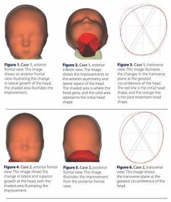
Case 1
A 4.5-month-old male presented with plagiocephaly secondary to torticollis and was a direct referral from the pediatrician. Treatment duration lasted 13 weeks. There was a 20mm increase in head circumference, and a 12mm improvement in CVA over the course of treatment. The cephalic index went from 93 percent to 92 percent. Excellent compliance was reported by the parents at each visit (wear time of 23 hours per day). Lateral and superior improvement was visible in the frontal plane. Inferior and superior transverse views showed significant improvement.
Case 2
A 4.5-month-old male born eight weeks prematurely presented with plagiocephaly secondary to torticollis, neonatal intensive care unit stay, and was a direct referral from the physical therapist. Treatment duration lasted seven weeks. Over the course of treatment, the patient’s head circumference increased 19mm, and CVA improved 10mm. The cephalic index actually increased from 84 percent to 86 percent due to the lateral expansion. Excellent compliance was reported by the parents at each visit (wear time of 23 hours per day). Lateral and superior improvement was visible in the frontal plane. Inferior and superior transverse views showed significant improvement.
The purpose of this case study is to illustrate the 3D changes of the head versus simply looking at crude point-to-point measurements. The design of both helmets was altered based on the parents’ primary concerns of lateral asymmetry. The superior opening of the helmet was shifted laterally to maintain contact on the superior aspect of the affected plates while also allowing sufficient relief for the entire flattened area of the head (see Figure 7, available in the digital issue). Further attention should be paid to the 3D presentation of plagiocephaly, and
a systematic way of reporting the associated outcomes should be established.
References
- Tiffany S. Glasgow, Faizi Siddiqi, Charles Hoff, Paul C. Young. Deformational plagiocephaly: development of an objective measure and determination of its prevalence in primary care. J Craniofac Surg. 2007; 18(1):85–92.
- Louis Argenta, Lisa David, James Thompson. Clinical classification of positional plagiocephaly. J Craniofac Surg 2004 15;(3)68–372
- Mi-Hyang Han, Jin Young Kang, Hye Young Han, Yun-Hwa Cho, Dae-Hyun Jang. Relationship between starting age of cranial-remolding-orthosis therapy and effectiveness of treatment in children with deformational plagiocephaly. Childs Nerv Syst. 2017;33(8):1349-1356.
- Tiffany Graham, Kelly Millay, Jijia Wang, Beverley Adams-Huet, Elizabeth O’Briant, Madison Oldham, Shacoya Smith. Significant factors in cranial remolding orthotic treatment of asymmetrical brachycephaly. J Clin Med. 2020;9(4):1027.
- Sybill D. Naidoo, Gary B. Skolnick, Anthony D. Galli Jr, Kamlesh B. Patel. (2019) Head shape retention following helmet therapy for deformational plagiocephaly. J Craniofac Surg. 2019;(6):1842-1844.
- Renske M. van Wijk, Leo A. van Vlimmeren, Catharina G. M. Groothuis-Oudshoorn, Catharina P. B. Van der Ploeg, Maarten J. Ijzerman, Magda M. Boere-Boonekamp. Helmet therapy in infants with positional skull deformation: randomised controlled trial. BMJ. 2014;348:g2741.
- Lynne B. Hutchison, Luke A.D. Hutchison, John M.D. Thompson, Ed A. Mitchell. Quantification of plagiocephaly and brachycephaly in infants using a digital photographic technique. Cleft Palate Craniofac J. 2005;42(5):539-547.
- James T. Thompson, Lisa R. David, Benjamin Wood, Anne Argenta, Jordan Simpson, Louis C. Argenta. Outcome analysis of helmet therapy for positional plagiocephaly using a three-dimensional surface scanning laser. J Craniofac Surg. 2009;20(2):362-365.
- Helena Sophie Visse, Ulrich Meyer, Christoph Runte, Holger Maas, Dieter Dirksen. Assessment of facial and cranial symmetry in infants with deformational plagiocephaly undergoing molding helmet therapy. J Craniomaxillofac Surg. 2020;48(6):548-554.
- Angelo B. Lipira, Shayna Gordon, Tron A. Darvann, Nuno V. Hermann, Andrea E. Van Pelt, Sybill D. Naidoo, Daniel Govier, Alex A. Kane. Helmet versus active repositioning for plagiocephaly: a three-dimensional analysis. Pediatrics. 2010;126(4):e936-945.

