Researchers at Brown University (Brown), Providence, Rhode Island, have created nanoscale surfaces for implanted materials that mimic the contours of natural skin, according to a university press release. The surfaces attract skin cells that, over time, are shown to build a natural seal against bacterial invasion. The group also created a molecular chain that allows an implant surface to be covered with skin cell-growing proteins, further accelerating skin growth. The findings have implications for implantable prostheses, for which the risk of infection at the skin/implant interface has been of concern. The results are published in the April 2011 Journal of Biomedical Materials Research A.
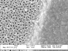
Nanotubular surfaces. Anodizing the titanium surface of a surgical implant, left, yields a roughened surface of nanotubes, which skin cells then colonize more quickly. Photograph courtesy of Webster Lab/Brown University.
“You need to close (the area) where the bacteria would enter the body, and that’s where the skin is,” said Thomas Webster, PhD, associate professor of engineering and orthopedic surgery at Brown.
Webster and the Brown research team report two ways in which they modified the surface of titanium leg implants to promote skin-cell growth, thereby creating a natural skin layer and sealing the gap where the device has been implanted into the body. The researchers also created a molecular chain to sprinkle skin-growing proteins on the implant to hasten skin growth.
The researchers, including Melanie Zile, a Boston University student who worked in Webster’s lab as part of Brown’s Undergraduate Teaching and Research Awards program, and Sabrina Puckett, PhD, who earned her engineering doctorate last May, created two different surfaces at the nanoscale, dimensions less than a billionth of a meter.
In the first approach, scientists fired an electron beam of titanium coating at the abutment (the piece of the implant inserted into the bone), creating a landscape of 20-nanometer mounds. These mounds imitate the contours of natural skin and trick skin cells into colonizing the surface and growing additional keratinocytes, or skin cells.
Webster knew such a surface, roughened at the nanoscale, worked for regrowing bone cells and cartilage cells, but was unsure whether it would be successful at growing skin cells. This may be the first time that a nanosurface created this way on titanium has been shown to attract skin cells.
The second approach, called anodization, involved dipping the abutment into hydrofluoric acid and then giving it a jolt of electric current. This causes the titanium atoms on the abutment’s surface to scatter and then regroup as hollow, tubular structures rising perpendicularly from the abutment’s surface. As with the nanomounds, skin cells quickly colonize the nanotubular surface.
In laboratory (in vitro) tests, the researchers report nearly a doubling of skin-cell density on the implant surface; within five days, the keratinocyte density reached the point at which an impermeable skin layer bridging the abutment and the body had been created.
“You definitely have a complete layer of skin,” Webster said. “There’s no more gap for the bacteria to go through.”
To further promote skin-cell growth around the implant, Webster’s team looked to FGF-2, a protein secreted by the skin to enhance skin cell growth. Researchers came up with a synthetic molecular chain to bind FGF-2 to the titanium surface, while maintaining the protein’s skin-cell growing ability. In vitro tests showed the greatest density of skin cells on abutment surfaces using the nanomodified surfaces laced with FGF-2. Moreover, the nanomodified surfaces create more surface area for FGF-2 proteins than would be available on traditional implants.
The next step is to perform in vivo studies; if they are successful, human trials could begin, although Webster said that could be years away.
The U.S. Department of Veterans Affairs and the U.S. National Science Foundation funded the research.
Editor’s note: This story has been adapted from materials provided by Brown University.